3D virtual histopathology for precision medicine
Topical lead: PD Dr. R. Zboray

We developed novel, clinically applicable 3D virtual histopathology methods for different tumor types (thyroid, colorectal) using phase-contrast micro- and nano-CT methods . We combine imaging-based biomarker discovery with molecular (multi-omics) analysis to get a holistic approach to tumor diagnostic and patient management and prognosis. Furthermore, we apply radiomics methods in a novel approach on micro-CT of tissue micro arrays to couple underlying genetic mutations and other patient specific information with microscale tumor textures. We provide a major contribution to the establishment and development of the Swiss PHRT Imaging HUB.
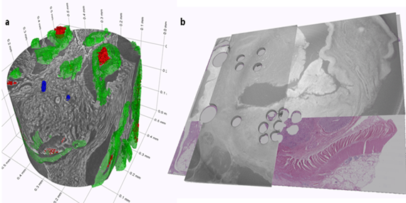
Figure caption: High-resolution phase-contrast nanoCT of a punch out core (600 micron diameter) from a whole biopsy block of colorectal tumor showing colonic crypts-green, necrotic cancer cell clusters- red and small tumor cells clusters (tumor budding) – blue (left). Registration of a H&E topslice with the 3D X-ray histopathology of a whole colorectal tumor biopsy block (right).
- K. Tajbakhsh, A Neels, Elena Fadeeva, J. C. Larson, O. Stanowska, A. Perren, R. Zboray. A comprehensive study of micro-CT for 3D virtual histology of FFPE tissue blocks. IEEE Access, vol. 12, pp. 78304-78316, (2024) https://doi.org/10.1109/ACCESS.2024.3407733
- K. Tajbakhsh, O Stanowska, A. Neels, A. Perren, R. Zboray (2024). 3D virtual histopathology by phase-contrast X-ray micro-CT for follicular thyroid neoplasms. IEEE Trans Med Imaging. Volume: 43, Issue: 7, July 2024
-
Share
