Blood microclots imaging
Topical lead: Dr. Peter Nirmalraj

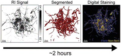
The coronavirus disease 2019 (COVID-19) has impacted health globally. Cumulative evidence points to long-term effects of COVID-19, such as cardiovascular and cognitive disorders, diagnosed in patients even after the recovery period. Micrometer-sized blood clots and hyperactivated platelets are potential indicators of long-term COVID. Using 3D digital holo-tomographic microscopy (DHTM), we show that we can resolve microclot structures in the plasma of patients with different sub phenotypes of COVID-19 in a label-free manner. Based on 3D refractive index (RI) tomograms, the size, dry mass, and prevalence of microclot composites can be quantified and parametrically differentiated from fibrin-rich microclots and platelet aggregates in the plasma of COVID-19 patients. Our imaging and analysis workflow is built around a commercially available DHT microscope capable of operation in clinical settings with a two-hour time period from sample preparation and data acquisition to results.
Figure caption: Imaging microclots in blood using 3D holotomography to identify the local chemical composition of the clots based on differences in refractive index values.
Reference:
- 3D holo-tomographic mapping of COVID-19 microclots in blood to assess disease severity, Bergaglio, T., Synhaivska, O., & Nirmalraj, P. N., Chemical & biomedical imaging. (2024), https://doi.org/10.1021/cbmi.3c00126
-
Share
