Bone structure and composition imaging
Topical lead: Dr. J. Schwiedrzik, Prof. Dr. A. Neels

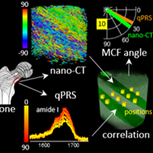
We combine complementary imaging techniques to study the structure and composition of the human bone. Organic and inorganic bone constituents can be simultaneously imaged in 3D with nanoscale resolution using nano-CT data and Raman spectroscopy.
Using machine-learning segmentation of nano-CT data, orientation of mineralized collagen fibrils could be quantified and correlated to quantitative polarized Raman spectroscopic measurements. We explore the observed structural and compositional variations in human cortical bone from the inferior region of the femoral neck.
Figure caption: Combining nano-CT and quantitative polarized Raman spectroscopy for determining the mineralization and orientation of bone tissue with a submicron spatial resolution.
References:
- Human Bone Ultrastructure in 3D: Multimodal Correlative Study Combining Nanoscale X ray Computed Tomography and Quantitative Polarized Raman Spectroscopy Kochetkova T et al., in submitted
- Combining polarized Raman spectroscopy and micropillar compression to study microscale structure-property relationships in mineralized tissues, Kochetkovaa T, Peruzzia C, Braun O, Overbeck J, Maurya A, Neels A, Calame M, Michler J, Zysset P, Schwiedrzik J; Acta Biomaterialia, 119, 390 (2021). https://doi.org/10.1016/j.actbio.2020.10.034
-
Share
