Dynamic Biplane Radiographic Imaging and Motion Analysis for Musculoskeletal Biodynamics Research
Topical lead: Prof. Dr. Ameet Aiyangar

Our long-term goal is to identify biomechanical markers and mechanical phenotypes of degenerative musculoskeletal joint disorders from multi-modality in vivo imaging, and exploit this knowledge to enable appropriate, patient-specific diagnoses and treatment strategies. To gain fundamental understanding of normal joint function, and, correspondingly, the aberrations due to injury or disease, not only is it essential to capture the gross rotational movements such as flexion, extension, abduction and adduction—osteokinematics—, but also the finer rolling and sliding movements—arthrokinematics, which requires measurement systems with high fidelity and sub-millimeter accuracy.
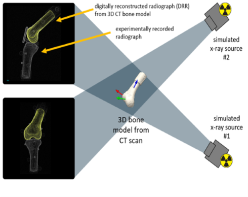
We achieve this by recording high-speed x-ray videos of bone motion without skin motion artifacts simultaneously in two directions during a functional movement with the help of a high frame rate Dynamic Biplane Radiographic Imaging system. The acquired images are then tracked non-invasively (i.e. without implanted beads) with a model-based bone tracking system to obtain three-dimensional joint kinematics including translational and rotational components with sub-millimeter accuracy. One project (Swiss National Science Foundation 10.003.251) aims to identify and quantify novel biomechanical markers based on a dynamic mechanical phenotype of unstable lumbar spine motion in patients with lumbar spinal stenosis with concomitant spondylolisthesis to enable enhanced decision making in the management of the disease. A second project (Swiss National Science Foundation 10.000.342) involves understanding the role of bone loss in recurrent shoulder instability. The work is a collaborative effort with clinicians from Inselspital University Hospital, Bern.
Figure caption: Marker less model-based bone tracking system.
-
Share
