X-ray analytics in motion
Dynamic 3D images for strong shoulders
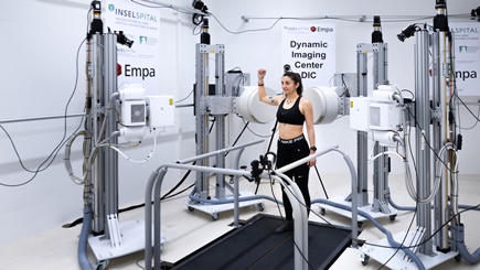
Shoulder instabilities are difficult to diagnose as they usually only occur when the shoulder joint is in motion. A time-resolved 3D analysis now makes it possible for the first time to precisely capture these dynamics. Empa researcher Ameet Aiyangar combines X-ray videos with virtual 3D models of the joints to detect these instabilities with millimeter precision.

After a shoulder injury has been treated, patients are often left with a feeling of insecurity – many of them report that their shoulder “doesn't hold” or “slips out easily”. When diagnosing shoulder instabilities, doctors often have to rely on these subjective assessments. The reason: Conventional imaging methods do not capture the movement of the shoulder. In contrast, the newly developed 4D analysis by Empa researcher Ameet Aiyangar goes beyond static imaging: “We combine high-precision X-ray videos from two perspectives and use them to reconstruct a four-dimensional motion analysis – in other words, a 3D image while the shoulder is moving.”
Optimized therapy instead of unnecessary surgery

The shoulder is the most mobile joint in the human body and is therefore particularly susceptible to injury. It is true that only around two percent of people dislocate their shoulder joint at some point in their lives. After treatment, however, many patients return to the clinic because they continue to have complaints or are dissatisfied with the result. According to Aiyangar, every other affected person suffers another dislocation of the shoulder joint. “This is particularly problematic with surgical procedures – up to two thirds of patients who have undergone surgery dislocate their shoulder again and come back to seek help. This shows that there is great potential for optimization in diagnostics and treatment planning.”
Currently, the stability of the shoulder joint is tested manually – a method whose accuracy heavily depends on the experience of the physician or physiotherapist. Static imaging procedures such as X-rays, magnetic resonance imaging (MRI) or computer tomography (CT) are only useful as a supplement, as they do not capture dynamic movement sequences. With the new technology, the Empa researchers can quantitatively measure and specifically analyze instabilities for the first time. This provides doctors with a more precise basis for deciding whether physiotherapeutic treatment is sufficient or whether surgery is necessary. “This avoids unnecessary surgical interventions or does not unnecessarily delay sensible ones, thus enabling individually optimized therapy,” says Aiyangar.
Measuring shoulder instabilities with millimeter precision
A biplanar radiographic imaging system (DBRI) installed at sitem-insel, the Swiss Institute for Translational and Entrepreneurial Medicine in Bern, is used for the dynamic 3D images of the shoulder. It was developed in close collaboration with Empa and Inselspital Bern. First, the test subjects perform specific shoulder movements, which are recorded from two different perspectives using synchronized X-ray images. Additional CT scans are used to reconstruct detailed 3D models of the bones with anatomical landmarks. A tracking procedure based on this determines the exact position of the shoulder joint. Finally, optimization software calculates the movement sequences of the shoulder.
This enables the researchers to record not only the rough movement patterns of a joint, but also the smallest rolling and sliding movements that are crucial for stability – with an accuracy in the range of 0.1 to 0.5 millimeters. “This is a decisive step forward, because conventional motion sensors with infrared cameras and markers on the skin are inaccurate by 20 to 40 millimeters – far too imprecise to reliably detect instabilities,” says Aiyangar.
Aiming for diagnosis without radiation-based imaging
The next step will be a joint study with Inselspital in Bern, with which there is a long-standing research collaboration. Around 40 patients with untreated shoulder instability are being sought to be examined before and after targeted muscle strengthening. Individual musculoskeletal models will play a central role to analyze the interactions between muscles, joints and forces. “In the long term, we hope that our four-dimensional movement analysis will find its way into clinical practice. This is because the problem with treating joint instability lies mainly in the dynamics,” explains Aiyangar. Based on the biomechanical study data, the Empa researcher also wants to develop force-controlled kinematic models that take into account muscle forces and joint loads in addition to movement. In future, these should enable a patient-specific dynamic analysis of shoulder instability – and thus a comprehensive clinical diagnosis without radiation-based imaging.
Prof. Dr. Ameet Aiyangar
Mechanical Systems Engineering
Phone +41 58 765 4508
Medical technology
Focusing on people: In the Research Focus Area Health, Empa researchers are developing pioneering solutions for the medicine of tomorrow – precisely where “conventional” materials meet living ones, i.e. cells and tissue. There are polymers that light up when there is an infection with certain germs, tiny gold particles against cancer or “nanozymes” that help mothers with complications during pregnancy without harming the foetus – and much more.
Read the latest EmpaQuarterly online or download the PDF version.
-
Share


