Minimally invasive cancer therapy
A new "gold" standard against metastases
If tumors spread in the abdomen, it is difficult to detect and fight these metastases. Researchers at Empa, ETH Zurich and the University of Zurich are therefore developing an efficient and gentle therapy using gold nanoparticles. Applied directly into the abdominal cavity, the gold particles are designed to detect tumor cells, penetrate them and kill them with heat.

If organs in the abdomen are affected by cancer, tumor cells can spread into the abdominal cavity from affected organs such as the bowel or the ovaries, for example. Despite medical advances, these metastases in the peritoneum are still difficult to diagnose and combat. The chances of recovery are thus rather small, as successful treatment depends on being able to detect and remove every cancerous cell. A team of researchers from Empa, ETH Zurich and the University of Zurich is therefore developing nanoparticles made of gold that specifically attack and eliminate metastases in the peritoneum. Initial laboratory results on the efficient and gentle treatment, which is applied directly into the abdominal cavity, are promising.
Trojan horse against cancer cells

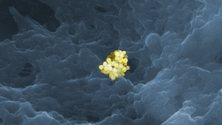
The researchers chose a highly biocompatible material as the starting material for the nanomedical cancer therapy: gold. If, however, the precious metal can be introduced into cancer cells, very much like a Trojan horse, it turns out to be a highly effective weapon. As gold particles absorb light from the infrared (IR) range and convert it into heat, they can destroy the tumor from the inside using hyperthermia.
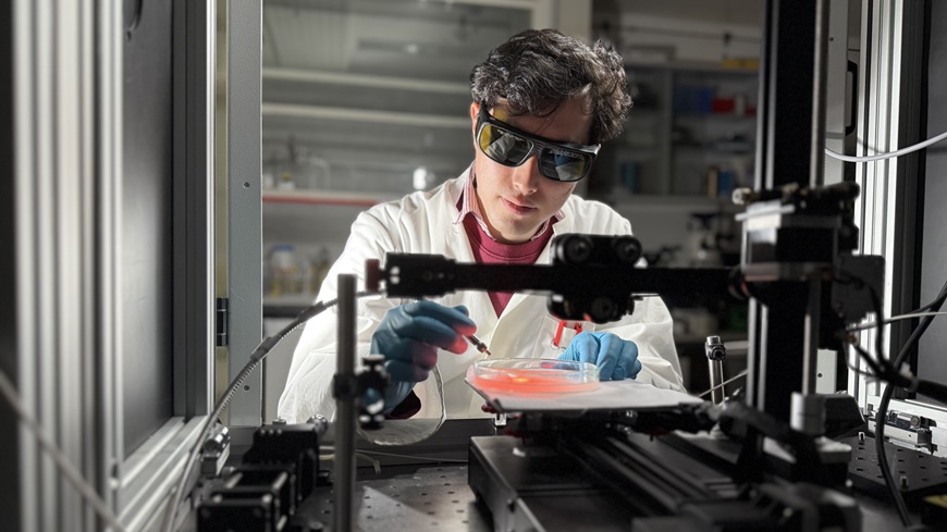
The team led by Inge Herrman from the laboratories of Empa, ETH Zurich and Balgrist University Hospital is developing and analyzing the nanoparticles together with the Department of Visceral and Transplant Surgery at the University Hospital Zurich and the University of Fribourg. The aim of the project is to synthesize nanoparticles that effectively and gently eliminate metastases.
The researchers are taking a new approach to delivering the nanomedicine to the site of action – i.e. the tumor: While conventional therapies are administered as an infusion via a drip, the nanoparticles are to be applied directly into the abdominal cavity during a minimally invasive procedure in order to increase efficiency. This also makes it possible to reduce the amount of chemotherapy drugs administered at the same time, which should reduce harmful side effects. “In laboratory experiments with tissue samples, we have already been able to show that the direct administration of gold nanoparticles into the abdominal cavity leads to efficient and selective uptake into the desired tissue,” says Empa researcher Oscar Cipolato from Empa's Nanomaterials in Health laboratory in St. Gallen. To further improve this, the researchers are now optimizing the shape, surface structure and size of the nanoparticles. Further experiments are currently underway to determine whether gold spheres up to 400 nanometers in size or gold rods just a few nanometers thick are better suited for this purpose.
A sleuth made of pure gold

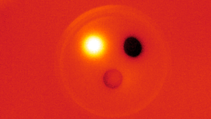
To ensure that only metastatic tissue heats up to over 42 degrees when patients are irradiated with near-IR light, the nanomedicines are to be equipped with antibodies against tumor tissue. In this way, the nanoparticles exclusively detect cancer cells. Such a customized therapy is therefore less stressful for the patient, as less surrounding healthy tissue is damaged.
The nanogold-based combination therapy should be used in everyday clinical practice as soon as possible. This requires basic information on the effect of nanoparticles and their distribution within tumors and within the human body. To close this gap, the researchers have optimized imaging techniques so that nanomaterials can be analyzed at the level of individual cells. Thanks to a combination of light and electron microscopy, they can precisely determine the interaction of the nanoparticles with the cells and their distribution in the tissue. The researchers hope that this better understanding of nanoparticles will help to drive forward new developments in nanomedicine for more effective and gentle cancer treatment.
Prof. Dr. Inge Herrmann
Particles-Biology Interactions Lab
Tel. +41 58 765 71 53
ETH Nanoparticle Systems Engineering Laboratory
Oscar Cipolato
Empa Particles-Biology Interactions
Tel. +41 58 765 7699
Wound soldering with nanothermometers
This study is funded by the SNSF, Swiss National Science Foundation (Eccellenza grant No. 181290) and by the Promedica Foundation.
O Cipolato, M Fauconneau, PJ LeValley, R Nisler, B Suter, IK Herrmann; An Augmented Reality Visor for Intraoperative Visualization, Guidance, and Temperature Monitoring Using Fluorescence; Journal of Biophotonics (2024); doi: 10.1002/jbio.202400417
Medical technology
Focusing on people: In the Research Focus Area Health, Empa researchers are developing pioneering solutions for the medicine of tomorrow – precisely where “conventional” materials meet living ones, i.e. cells and tissue. There are polymers that light up when there is an infection with certain germs, tiny gold particles against cancer or “nanozymes” that help mothers with complications during pregnancy without harming the foetus – and much more.
Read the latest EmpaQuarterly online or download the PDF version.
-
Share


