| |
| Das
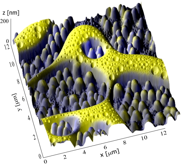
Resultat einer kombinierten, dreidimensionalen
ToF-SIMS-/SFM-Oberflächenanalyse einer
PCBM/CyI-Polymermischung, die in der Empa-Abteilung
«Funktionspolymere» zur Herstellung organischer
Solarzellen verwendet wird. |
| |
| Was
haben ein Pinguin und die Oberfläche einer Solarzelle
miteinander zu tun? Nicht viel, gibt Empa-Physikerin Laetitia
Bernard zu. Doch hätte sie schmunzeln müssen, als sich
bei der Bearbeitung des Abbilds einer Polymermischung, die für
die Herstellung von neuartigen organischen Solarzellen
benötigt wird, an einer bestimmten Stelle immer deutlicher die
Umrisse eines Pinguins herausschälten. Ein kleines Detail in
der komplexen Welt der Hochleistungsmikroskopie. Das an der Empa
entwickelte 3D-NanoChemiscope bildet Proben nicht nur
nanometergenau ab, sondern kann auch erstmalig präzis
Aufschluss darüber geben, welche chemischen Elemente sich in
einer Probe wo angeordnet haben. Simultan und dreidimensional
lassen sich damit mechanische Eigenschaften wie Härte,
Elastizität oder Reibungskoeffizient, aber auch die chemischen
Eigenschaften von Oberflächenstrukturen bestimmen. Im Falle
des «Pinguin»-Bildes heisst das: das 3D-NanoChemiscope
erfasst nicht nur die Umrisse des «Pinguins», sondern
deckt auch auf, welche Polymere sich auf seinem
«Schnabel», auf seinem «Auge» und «um
ihn herum» angesiedelt haben. Mithilfe dieser Analyse
können die SolarzellenforscherInnen effizient die Mechanismen
ihrer Materialien kontrollieren und entsprechend Zusammensetzung
oder Konzentration ihrer Polymermischung ändern. So lassen
sich neue Strukturen und somit auch andere Eigenschaften für
die Solarzelle herbeiführen. |
| |
|
| |
|
|
|
Einige der
vielen einzelnen Bilder, aus denen das 3D-NanoChemiscope die
3D-Ansicht generierte. Mit dem SFM wird die Oberfläche
topographisch abgebildet (Das Bild links zeigt einen 12µm x
12µm grossen Ausschnitt. Die im Bild sichtbaren
Höhenunterschiede betragen 100-200nm). Mit dem TOF-SIMS kann
gezeigt werden, wo sich die verschiedenen Stoffe, bzw. Polymere der
Polymermischung auf der Oberfläche angesiedelt haben (Bilder
Mitte und rechts zeigen C-+C2- und CN-+I-
Ionen). |
| |
|
| |
Rasterkraftmikroskop und High-End-Massenspektrometer
Möglich macht diese Analyse das 3D-NanoChemiscope,
das zwei bisherig unabhängige Techniken vereint. Das
Rasterkraftmikroskop (SFM, von engl. «scanning force
microscope») rastert mit einer ultrafeinen Nadel die
Oberfläche ab, das High-End-Massenspektrometer (ToF-SIMS, von
engl. «time-of-flight secondary ion mass spectrometry»)
ermittelt, aus welchen Stoffen sich die oberste monomolekulare
Schicht der Festkörperoberfläche zusammensetzt, indem
Ionenstrahlen das Material «beschiessen». |
| |
| Wollte
man bis anhin Oberflächen sowohl auf chemische als auch auf
physikalische Eigenschaften untersuchen, so musste die Probe in
zwei verschiedenen Geräte analysiert werden. Doch durch das
Hin- und Hertragen vom einen zum anderen Gerät lief man immer
Gefahr, die Probe zu verschmutzen oder zu oxidieren. Ausserdem war
es praktisch unmöglich, die im SFM untersuchte Stelle exakt
wiederzufinden. Was lag also näher, als die beiden Geräte
zu «vereinen»? Damit stiessen die Physikerinnen und
Maschinenentwickler bis anhin an ihre Grenzen. In einem
vierjährigen, von der EU geförderten Projekt entwickelte
Projektleiterin Laetitia Bernard mit Empa-ForscherInnen und
Partnern aus Akademie und Industrie in akribischer Arbeit ein neues
Gerät, in dem ein SFM und ein ToF-SIMS in einer
Ultrahochvakuumkammer möglichst nah nebeneinander angeordnet
sind. |
| |
|
| |
|
|
|
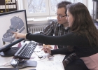
Sasa
Vranjkovic, Maschinenbauer, und Laetitia
Bernard, Leiterin des Projekts 3D-NanoChemiscope, diskutieren
den Konstruktionsplan eines Bauteils. |
| |
|
| |
| Die
MikroskopexpertInnen rüsteten das 3D-NanoChemiscope dazu mit
einem neuartigen, eigens entwickelten Transportsystem aus, das die
Probe auf einer Schiene mit einer Beschichtung aus
diamantähnlichem Kohlenstoff (DLC) mittels Piezomotor sanft
hin- und herschiebt. Der Probenhalter kann Bewegungen auf fünf
Achsen ausführen, sodass sich die zu untersuchende Stelle aus
den unterschiedlichsten Positionen analysieren lässt. |
| |
| Nach
seiner Fertigstellung steht der Prototyp – ein Ungetüm
aus glänzendem Aluminium, 1 Meter lang, 70 Zentimeter breit
und an die 1 Meter 70 hoch – nun beim Projektpartner ION-TOF
GmbH im deutschen Münster im Einsatz und wird von
Industriekunden und Forschungspartnern genutzt. Der Bau weiterer
Geräte steht an, Kunden zeigen sich sehr interessiert und sind
bereit, Beträge im siebenstelligen Frankenbereich dafür
zu bezahlen. |
| |
| |


